

Drug Information
- Classification of drug : Ethical drug
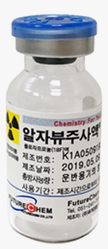
- Shape and form : This drug is packed in a lead shielding container. It is colorless liquid without foreign substances packed in colorless transparent vial.
- Effective component : 18florapronol liquid
- Item Standard Code : 201800667
Efficacy
This drug is used for Positron Emission Tomography (PET) in the following cases.
It is used for auxiliary diagnosis of clinical test by estimating the presence of beta amyloid neuritic plaque in the brain of adult cognitive disorder patents who need to be tested for Alzheimer’s disease or the cause of cognitive disorder.
The negative scan in PET makes it possible to estimate that there is less or no beta amyloid neuritic plaque while positive scan in PET makes it possible to estimate that there is beta amyloid neuritic plaque. (Refer to the usage & dose for the criteria for the judgment of negative scan and positive scan.) It does not necessarily correspond with the neuropathologic diagnosis of Alzheimer’s disease.
The sensitivity and specificity of the PET image obtained with this drug were evaluated on the basis of clinical diagnosis.
Beta amyloid neuritic plaque may exist in the elderly people who have normal cognition ability or those who have Alzheimer’s disease or other types of neurological disorder (dementia with Lewy bodies, dementia by Parkinson’s diseases, etc.), too. Thus, this drug should be used for supplementary diagnosis of other diagnostic testing.
As beta amyloid neuritic plaque in the grey matter may exist in the elderly who have no symptoms and the patients of Alzheimer’s disease or other types of neurological disorder (dementia with Lewy bodies, dementia by Parkinson’s diseases, etc.), it is not possible to diagnose Alzheimer’s disease or other types of cognition disorder independently.
The safety and efficacy of this drug have not been established for:

			- Predicting development of dementia or other neurological condition
			- Monitoring responses to therapies
It is not possible to predict the reaction to the materials other than amyloid neuritic plaque such as tau protein.
This drug is used for Positron Emission Tomography (PET) in the following cases. It is used for auxiliary diagnosis of clinical test by estimating the presence of beta amyloid neuritic plaque in the brain of adult cognitive disorder patents who need to be tested for Alzheimer’s disease or the cause of cognitive disorder. The negative scan in PET makes it possible to estimate that there is less or no beta amyloid neuritic plaque while positive scan in PET makes it possible to estimate that there is beta amyloid neuritic plaque. (Refer to the usage & dose for the criteria for the judgment of negative scan and positive scan.) It does not necessarily correspond with the neuropathologic diagnosis of Alzheimer’s disease. The sensitivity and specificity of the PET image obtained with this drug were evaluated on the basis of clinical diagnosis. Beta amyloid neuritic plaque may exist in the elderly people who have normal cognition ability or those who have Alzheimer’s disease or other types of neurological disorder (dementia with Lewy bodies, dementia by Parkinson’s diseases, etc.), too. Thus, this drug should be used for supplementary diagnosis of other diagnostic testing. As beta amyloid neuritic plaque in the grey matter may exist in the elderly who have no symptoms and the patients of Alzheimer’s disease or other types of neurological disorder (dementia with Lewy bodies, dementia by Parkinson’s diseases, etc.), it is not possible to diagnose Alzheimer’s disease or other types of cognition disorder independently. The safety and efficacy of this drug have not been established for: - Predicting development of dementia or other neurological condition - Monitoring responses to therapies It is not possible to predict the reaction to the materials other than amyloid neuritic plaque such as tau protein.
Detailed information
Usage & Dose
A clinician who has the experience of treating neurodegenerative disease should perform PET scan using this drug.
The image obtained with this drug refers to the interpretation of PET image using [18F]florappronol, and it should be read and interpreted by only trained experts. Radiopharmaceuticals including this drug should be used only by or under the supervision of a medical doctor who was trained in an educational course approved by the government agency authorized to permit the use of radionuclides and has related experience (the experience in safe use and handling of radionuclides).
A clinician who has the experience of treating neurodegenerative disease should perform PET scan using this drug. The image obtained with this drug refers to the interpretation of PET image using [18F]florappronol, and it should be read and interpreted by only trained experts. Radiopharmaceuticals including this drug should be used only by or under the supervision of a medical doctor who was trained in an educational course approved by the government agency authorized to permit the use of radionuclides and has related experience (the experience in safe use and handling of radionuclides).
Dosage
Intravenous injection of the dosage corresponding with [18F] florappronol 370 MBq(10 mCi).
Administration method
This drug shall be administered once by intravenous injection. The optimal administration speed, the limit of safe dosage and the standard of safety in multiple administration have not been established. Flushing with 5~10 mL of physiological saline (9 mg/mL, 0.9%) shall be done after administration of the drug. If the dosage of this drug is 0.5 ~ 1 mL, use a syringe of about 1 mL capacity and flushing shall be done with physiological saline.
This drug shall be injected into the vein without fail in order to prevent extravasation of the drug.
The dosage for the last patient (who is administered the drug long after its production time) shall be calculated based on the end of synthesis (EOS) using decay constant.
This drug shall not be diluted.
This drug shall be administered once by intravenous injection. The optimal administration speed, the limit of safe dosage and the standard of safety in multiple administration have not been established. Flushing with 5~10 mL of physiological saline (9 mg/mL, 0.9%) shall be done after administration of the drug. If the dosage of this drug is 0.5 ~ 1 mL, use a syringe of about 1 mL capacity and flushing shall be done with physiological saline. This drug shall be injected into the vein without fail in order to prevent extravasation of the drug. The dosage for the last patient (who is administered the drug long after its production time) shall be calculated based on the end of synthesis (EOS) using decay constant. This drug shall not be diluted.
Imaging Acquisition
Acquire the PET/CT image for 30 minutes from 30 minutes after intravenous injection of about 370 MBq(10 mCi) of the drug.
			PET Image Display
			1) the image at the workstation exclusive for reading.
			2) Display of the image shall be done on the basis of axial image referring to coronal image and sagittal image.
			3) In the sagittal image, set the anterior commissure-posterior commissure line (AC-PC line) horizontally.
			4) Set the axial image and the coronal image to make both cerebral hemispheres symmetrical.
			5) Display the color scale spectrum using the standard scale and referring to other color scales (Ex: rainbow, hot metal, etc.).

            All the color scales including spectrum shall satisfy all of the following 3 condition. Another color scale may be chosen if the spectrum does not satisfy this standard.
            It is possible to refer to a single-color scale (ex: grey scale) in addition to the above color scales.
            a. It should be able to discriminate the uptake area which is in a higher or lower intensity than the pons.
            b. Even at an area with no or very less uptake of amyloid such as the cerebellum, it should be able to check the color image with naked eye by adjusting the intensity.
            c. It should be able to divide the areas showing more than 50% of the maximum uptake into 5 or more colors according to intensity.
Acquire the PET/CT image for 30 minutes from 30 minutes after intravenous injection of about 370 MBq(10 mCi) of the drug.
PET Image Display
1) the image at the workstation exclusive for reading.
2) Display of the image shall be done on the basis of axial image referring to coronal image and sagittal image.
3) In the sagittal image, set the anterior commissure-posterior commissure line (AC-PC line) horizontally.
4) Set the axial image and the coronal image to make both cerebral hemispheres symmetrical.
5) Display the color scale spectrum using the standard scale and referring to other color scales (Ex: rainbow, hot metal, etc.).
All the color scales including spectrum shall satisfy all of the following 3 condition. Another color scale may be chosen if the spectrum does not satisfy this standard.
It is possible to refer to a single-color scale (ex: grey scale) in addition to the above color scales.
a. It should be able to discriminate the uptake area which is in a higher or lower intensity than the pons.
b. Even at an area with no or very less uptake of amyloid such as the cerebellum, it should be able to check the color image with naked eye by adjusting the intensity.
c. It should be able to divide the areas showing more than 50% of the maximum uptake into 5 or more colors according to intensity.
Image Interpretation
The image acquired by using this drug refer to the interpretation of the PET image using [18F]florappronol, and it should be interpreted by only trained experts. The purpose of interpreting the image obtained with this drug is not clinical diagnosis but to estimate the presence of brain beta amyloid neuritic plaque in the brain. Image interpretation is performed regardless of the clinical characteristic of the patient and it is recognizing the characteristic of the image.

				1) The evaluation of the positive and negative scan of the image is designated by the degree of uptake of radiopharmaceuticals at the cerebral cortical grey matter and white matter.
At this time, clinical information or absorption of signal by the cerebellum is not reflected to image interpretation.
				2) In axial image, the difference in the degree of uptake between the cerebral cortical grey matter and white matter is compared with naked eye going from the lower part to the upper part of the cerebrum and evaluate for a positive scan or a negative scan according to the following criteria.
-A negative scan is defined as a case in which the border between the grey matter and the white matter is observed as the degree of uptake by the grey matter is lower than the degree of uptake by the white matter in all areas of cerebral cortex.
However, a negative scan may have the following characteristics.

				a. The white matter tract connecting the frontal lobe and the parietal lobe is observed clearly. Or,
					b. The white matter tract connecting the occipital lobe and the temporal lobe is observed clearly. Or,
					c. The shape of finger is observed due to the uptake of the whiter matter of the frontal lobe.-A positive scan is defined as a case in which the uptake by one or more cerebral cortical grey matter is the same as or more than that of the adjacent white matter.
However, a positive scan may have the following characteristics.
a. It is difficult to observe the white matter tract connecting the frontal lobe and the parietal lobe. Or,
					b. It is difficult to observe the white matter tract connecting the occipital lobe and the temporal lobe. Or,
					c. The uptake of the grey matter by the medial parietal lobe (precuneus) has increased.
The image acquired by using this drug refer to the interpretation of the PET image using [18F]florappronol, and it should be interpreted by only trained experts. The purpose of interpreting the image obtained with this drug is not clinical diagnosis but to estimate the presence of brain beta amyloid neuritic plaque in the brain. Image interpretation is performed regardless of the clinical characteristic of the patient and it is recognizing the characteristic of the image. 1) The evaluation of the positive and negative scan of the image is designated by the degree of uptake of radiopharmaceuticals at the cerebral cortical grey matter and white matter. At this time, clinical information or absorption of signal by the cerebellum is not reflected to image interpretation. 2) In axial image, the difference in the degree of uptake between the cerebral cortical grey matter and white matter is compared with naked eye going from the lower part to the upper part of the cerebrum and evaluate for a positive scan or a negative scan according to the following criteria. -A negative scan is defined as a case in which the border between the grey matter and the white matter is observed as the degree of uptake by the grey matter is lower than the degree of uptake by the white matter in all areas of cerebral cortex. However, a negative scan may have the following characteristics.a. The white matter tract connecting the frontal lobe and the parietal lobe is observed clearly. Or, b. The white matter tract connecting the occipital lobe and the temporal lobe is observed clearly. Or, c. The shape of finger is observed due to the uptake of the whiter matter of the frontal lobe.-A positive scan is defined as a case in which the uptake by one or more cerebral cortical grey matter is the same as or more than that of the adjacent white matter. However, a positive scan may have the following characteristics.a. It is difficult to observe the white matter tract connecting the frontal lobe and the parietal lobe. Or, b. It is difficult to observe the white matter tract connecting the occipital lobe and the temporal lobe. Or, c. The uptake of the grey matter by the medial parietal lobe (precuneus) has increased.
Precautions
1. Do not administer the drug to the following patients
		1) This drug is contraindicated in patients with a history of hypersensitivity reaction to [18F]florapronol or excipient of this drug

		2. Adverse Reactions
		1) In the Phase 3 clinical trial conducted on 105 patients in total, 6 cases of adverse reaction occurred in 6 patients (5.71%). One case each of nausea, vomiting, strange walking, fever, pain and dizziness occurred and all were mild or medium level of adverse reaction. Any of the cases had cause-and-effect relationship with this drug and there was no serious adverse reaction.

		3. General Caution
            1) Administer the drug only when it is thought that the benefit of diagnosis is more than the disadvantage of exposure to radiation and use minimum dosage.
            2) As this drug is a radioactive product, handle it with care and pay attention to the protection of the safety of the patients and the medical staff who are exposed to radioactivity.
            3) Exposure to ionizing radiation may cause a cancer or genetic defect. The quantity of radioactivity used for the diagnosis of nuclear medicine is mostly less than 20mSv and there are few cases of adverse reaction.
            However, the use of the quantity of radioactivity for medical treatment may cause a cancer and mutation.
            4) Radiopharmaceuticals may be used by only those who have the license to use and handle radionuclides. Radiopharmaceuticals shall be received, stored, used, moved and discarded according to related laws and regulations.
            5) Radiopharmaceuticals shall be prepared by the user using the method which can satisfy radiological safety and pharmaceutical quality requirement.
            6) The patient shall be restricted from contacting infants and pregnant women for 24 hours after injection of the drug.
            7) This drug contains 10%(v/v) of ethanol and thus it shall be injected carefully to a patient who has hepatic failure or epilepsy.
            8) This drug contains sodium bisulfite and thus may cause such allergy as sulfurous acid anaphylaxis and some sensitive patients may suffer fatal or less serious asthmatic attack. While the comprehensive frequency of the sensitivity to sulfurous acid in ordinary people is not known, it seems that the sensitivity is low and those patients who are suffering from asthma showed more sensitivity to sulfurous acid.
            9) Risk for Image Misinterpretation and Other Errors: Errors may occur while using the PET image of this drug for examination of the presence of beta amyloid neuritic plaque in the brain.
            Image interpretation is conducted separately from the clinical information of the patient. The use of clinical information was not evaluated for interpretation of the image of this drug, and clinical information may cause an error in interpretation of the image of this drug.
            Noise made by movement may distort the image. The result of scan of this drug shows only the estimation of the presence of beta amyloid neuritic plaque in the brain at the time when the image is acquired, and the result of the negative scan does not rule out the possibility of revelation of brain beta amyloid neuritic plaque in the future.

		4. Drug Interaction
		Pharmacodynamic drug-drug interaction studies have not been performed in patients to establish the extent, if any, to which concomitant medications may alter [18F]florapronol image results.

		5. Administration for pregnant women and breast-feeding women
        1) Pregnant women: No clinical study was conducted on pregnant women. It is not known if injection of this drug for pregnant women may give harm to the fetus or fertility is not known.
        No reproduction-toxicity study was conducted on the toxicity of this drug for animals. All radiopharmaceuticals including this medicine has the possibility to give harm to the fetus. The possibility of causing damages to the fetus differs according to the stage of development of the fetus and the degree of dosage of the radiopharmaceuticals. This drug can be provided for pregnant women only when it is essential to do so. The women who have the possibility of pregnancy shall be checked for pregnancy before administration of this drug.
        2) Breast-feeding women: It is not known whether this drug is secreted through the mother’s milk. Many drugs are secreted through the milk and thus a breast-feeding mother shall avoid using this drug due to the risk of exposure of radioactivity to the baby or stop breast-feeding temporarily for 24 hours (more than 10 times of the half-life of the radioactivity decay of 18F isotope) after exposure to this drug.
        The mother shall be prohibited to contact the baby for 24 hours after injection of the drug.

		6. Administration for children
		The safety and efficacy of this drug in children have not been established.

		7. Administration for patients of liver failure and kidney failure
		The safety and efficacy of this drug in the patients with liver failure and kidney failure have not been established.

		8. Treatment at the time of administration of excessive quantity
        There is no report of the result of clinical study on administration of excessive quantity of this drug.
If this drug is administered excessively, supply water and recommend frequent urination and bowel movement in order to minimize the quantity of absorption of radiation by increasing the quantity of excretion of the patient. Be careful not to be contaminated by the radioactivity coming out of the patient.


		9. Precautions for storage and handling
		This drug is a radiopharmaceutical and it shall be put into a sealed container and stored at room temperature in a lead shielding container.

		10. Precautions for application
        1) This drug shall be injected into the vein without fail in order to prevent extravasation of the drug. (Refer to the usage & dose for the administration method.)
        2) When handling this drug, minimize the risk of exposure to radioactivity by the workers and other people by taking appropriate measures for safety through effective shielding, etc.

		11. Other
		1) Information for counseling with the patient
        ①In order to minimize the amount of absorption of radiation in the bladder, tell the patient to drink much water for several hours before and after the intravenous injection of this drug to be able to empty the bladder frequently, and secrete frequently for 24 hours after administration of the drug.
②In order to minimize the amount of exposure to radiation by the patient and the people around the patient, tell the patient to secrete using the same toilet for 12 hours after administration of the drug and flush the toilet several times after each time of secretion. After relieving oneself, the patient should wash hands thoroughly to prevent contamination by radioactivity.
If the clothes are contaminated by bold, urine or excrement, wash the clothes separately.

		12. Information for professionals

		1) Clinical Pharmacology
		This drug is a PET imaging tracer combining with the beta amyloid neuritic plaque which is known as the pathological cause of Alzheimer’s disease.
		① In vitro study
		The power of combination (IC50) with the beta amyloid neuritic plaque using the homogenate of the brain of an Alzheimer’s patient appeared to be 14 nM.
		② Ex vivo study
        An autoradiography research was conducted on Alzheimer’s disease patients and normal people in order to find out the selective labeling ability on amyloid neuritic plaque.
While the brain tissue of normal people did not combine with this drug, increase in combination of the brain tissue of Alzheimer’s disease patients with this drug was observed in areas with dense amyloid neuritic plaque in the brain of Alzheimer’s disease patients.

		2) Pharmacokinetics
		① Non-clinical Studies
        The absorption, distribution, metabolism and excretion of this drug were evaluated with mice. The drug was absorbed in the liver and kidney in a relatively high concentration in one minute after injection of the drug, and the concentration increased in the excrement organ of the bowel with the passage of time. The brain area showed a higher concentration immediately after the injection of the drug which disappeared quickly with the passage of time.
According to the results of metabolism study; the ratio of metabolome was low in the brain area but it increased relatively more in plasma with the passage of time and the ratio of metabolome increased in the liver from one minute after injection due to quick metabolism.
The major confirmed metabolome was in the form of phosphate made by phosphorylation of the hydroxyl in the isopropyl group.
The quantity of excretion was measured with the quantity of radioactivity until 4 hours after injection, and the quantity of excreted radioactivity was equivalent to about 15% of the dosage. Naturally, the rest quantity of radioactivity was kept in the urine and excrement remaining in the bladder and bowel before excretion.

		② Clinical Studies
        The clinical trial was conducted on a total of 10 subjects (5 normal subjects and 5 Alzheimer’s disease patients).
The 6 areas of interest (the frontal lobe, the temporal lobe, the parietal lobe, the occipital lobe, the white matter and the cerebellum) were set in the PET/CT image matching the MRI of the brain area. The standardized uptake value ratio (SUVR) was measured for each area of the brain in each time zone, and SUV was calculate based on the cerebellum.

As the indicators, the Tmax (the time taken for maximum uptake in the brain part) of cerebral cortex and the cerebellum, the Cmax(the standardized coefficient of maximum uptake in the brain part) and the T1/2(the time taken for reduction of Cmax into half) were calculate.

In all of the 10 subjects, the Tmax was less than 5 minute and the Cmax and T1/2 (min) in the 4 Alzheimer’s disease patients who showed uptake of this drug in their cerebral cortex were 2.54±0.43 and 16.63±10.74 respectively.
In the 5 normal subjects, the Cmax and T1/2 (min) of cerebral cortex were 3.0±0.61 and 8.55±1.64 respectively.

		3) Clinical Studies
		An open, single administration, blind method of the evaluator, Phase 3 clinical trial was conducted for a total of 105 subjects (53 Alzheimer’s disease patients, 16 non-Alzheimer’s disease patients and 36 normal subjects). The test drug was intravenously injected once with the quantity of 10 ± 1 mCi (370 ± 37 MBq). An independent evaluator conducted the evaluation.
    In the analysis of the PET image of this drug, the sensitivity of diagnosis was 90.6%(95% CI 82.7% ~ 98.4%) and the specificity of the diagnosis was 84.6%(95% CI 74.8% ~ 94.4%).
1. Do not administer the drug to the following patients
1) This drug is contraindicated in patients with a history of hypersensitivity reaction to [18F]florapronol or excipient of this drug
2. Adverse Reactions
1) In the Phase 3 clinical trial conducted on 105 patients in total, 6 cases of adverse reaction occurred in 6 patients (5.71%). One case each of nausea, vomiting, strange walking, fever, pain and dizziness occurred and all were mild or medium level of adverse reaction. Any of the cases had cause-and-effect relationship with this drug and there was no serious adverse reaction.
3. General Caution
1) Administer the drug only when it is thought that the benefit of diagnosis is more than the disadvantage of exposure to radiation and use minimum dosage.
2) As this drug is a radioactive product, handle it with care and pay attention to the protection of the safety of the patients and the medical staff who are exposed to radioactivity.
3) Exposure to ionizing radiation may cause a cancer or genetic defect. The quantity of radioactivity used for the diagnosis of nuclear medicine is mostly less than 20mSv and there are few cases of adverse reaction.
However, the use of the quantity of radioactivity for medical treatment may cause a cancer and mutation.
4) Radiopharmaceuticals may be used by only those who have the license to use and handle radionuclides. Radiopharmaceuticals shall be received, stored, used, moved and discarded according to related laws and regulations.
5) Radiopharmaceuticals shall be prepared by the user using the method which can satisfy radiological safety and pharmaceutical quality requirement.
6) The patient shall be restricted from contacting infants and pregnant women for 24 hours after injection of the drug.
7) This drug contains 10%(v/v) of ethanol and thus it shall be injected carefully to a patient who has hepatic failure or epilepsy.
8) This drug contains sodium bisulfite and thus may cause such allergy as sulfurous acid anaphylaxis and some sensitive patients may suffer fatal or less serious asthmatic attack. While the comprehensive frequency of the sensitivity to sulfurous acid in ordinary people is not known, it seems that the sensitivity is low and those patients who are suffering from asthma showed more sensitivity to sulfurous acid.
9) Risk for Image Misinterpretation and Other Errors: Errors may occur while using the PET image of this drug for examination of the presence of beta amyloid neuritic plaque in the brain.
Image interpretation is conducted separately from the clinical information of the patient. The use of clinical information was not evaluated for interpretation of the image of this drug, and clinical information may cause an error in interpretation of the image of this drug.
Noise made by movement may distort the image. The result of scan of this drug shows only the estimation of the presence of beta amyloid neuritic plaque in the brain at the time when the image is acquired, and the result of the negative scan does not rule out the possibility of revelation of brain beta amyloid neuritic plaque in the future.
4. Drug Interaction
Pharmacodynamic drug-drug interaction studies have not been performed in patients to establish the extent, if any, to which concomitant medications may alter [18F]florapronol image results.
5. Administration for pregnant women and breast-feeding women
1) Pregnant women: No clinical study was conducted on pregnant women. It is not known if injection of this drug for pregnant women may give harm to the fetus or fertility is not known.
No reproduction-toxicity study was conducted on the toxicity of this drug for animals. All radiopharmaceuticals including this medicine has the possibility to give harm to the fetus. The possibility of causing damages to the fetus differs according to the stage of development of the fetus and the degree of dosage of the radiopharmaceuticals. This drug can be provided for pregnant women only when it is essential to do so. The women who have the possibility of pregnancy shall be checked for pregnancy before administration of this drug.
2) Breast-feeding women: It is not known whether this drug is secreted through the mother’s milk. Many drugs are secreted through the milk and thus a breast-feeding mother shall avoid using this drug due to the risk of exposure of radioactivity to the baby or stop breast-feeding temporarily for 24 hours (more than 10 times of the half-life of the radioactivity decay of 18F isotope) after exposure to this drug.
The mother shall be prohibited to contact the baby for 24 hours after injection of the drug.
6. Administration for children
The safety and efficacy of this drug in children have not been established.
7. Administration for patients of liver failure and kidney failure
The safety and efficacy of this drug in the patients with liver failure and kidney failure have not been established.
8. Treatment at the time of administration of excessive quantity
There is no report of the result of clinical study on administration of excessive quantity of this drug.
If this drug is administered excessively, supply water and recommend frequent urination and bowel movement in order to minimize the quantity of absorption of radiation by increasing the quantity of excretion of the patient. Be careful not to be contaminated by the radioactivity coming out of the patient.
9. Precautions for storage and handling
This drug is a radiopharmaceutical and it shall be put into a sealed container and stored at room temperature in a lead shielding container.
10. Precautions for application
1) This drug shall be injected into the vein without fail in order to prevent extravasation of the drug. (Refer to the usage & dose for the administration method.)
2) When handling this drug, minimize the risk of exposure to radioactivity by the workers and other people by taking appropriate measures for safety through effective shielding, etc.
11. Other
1) Information for counseling with the patient
①In order to minimize the amount of absorption of radiation in the bladder, tell the patient to drink much water for several hours before and after the intravenous injection of this drug to be able to empty the bladder frequently, and secrete frequently for 24 hours after administration of the drug.
②In order to minimize the amount of exposure to radiation by the patient and the people around the patient, tell the patient to secrete using the same toilet for 12 hours after administration of the drug and flush the toilet several times after each time of secretion. After relieving oneself, the patient should wash hands thoroughly to prevent contamination by radioactivity.
If the clothes are contaminated by bold, urine or excrement, wash the clothes separately.
12. Information for professionals
1) Clinical Pharmacology
This drug is a PET imaging tracer combining with the beta amyloid neuritic plaque which is known as the pathological cause of Alzheimer’s disease.
① In vitro study
The power of combination (IC50) with the beta amyloid neuritic plaque using the homogenate of the brain of an Alzheimer’s patient appeared to be 14 nM.
② Ex vivo study
An autoradiography research was conducted on Alzheimer’s disease patients and normal people in order to find out the selective labeling ability on amyloid neuritic plaque.
While the brain tissue of normal people did not combine with this drug, increase in combination of the brain tissue of Alzheimer’s disease patients with this drug was observed in areas with dense amyloid neuritic plaque in the brain of Alzheimer’s disease patients.
2) Pharmacokinetics
① Non-clinical Studies
The absorption, distribution, metabolism and excretion of this drug were evaluated with mice. The drug was absorbed in the liver and kidney in a relatively high concentration in one minute after injection of the drug, and the concentration increased in the excrement organ of the bowel with the passage of time. The brain area showed a higher concentration immediately after the injection of the drug which disappeared quickly with the passage of time.
According to the results of metabolism study; the ratio of metabolome was low in the brain area but it increased relatively more in plasma with the passage of time and the ratio of metabolome increased in the liver from one minute after injection due to quick metabolism.
The major confirmed metabolome was in the form of phosphate made by phosphorylation of the hydroxyl in the isopropyl group.
The quantity of excretion was measured with the quantity of radioactivity until 4 hours after injection, and the quantity of excreted radioactivity was equivalent to about 15% of the dosage. Naturally, the rest quantity of radioactivity was kept in the urine and excrement remaining in the bladder and bowel before excretion.
② Clinical Studies
The clinical trial was conducted on a total of 10 subjects (5 normal subjects and 5 Alzheimer’s disease patients).
The 6 areas of interest (the frontal lobe, the temporal lobe, the parietal lobe, the occipital lobe, the white matter and the cerebellum) were set in the PET/CT image matching the MRI of the brain area. The standardized uptake value ratio (SUVR) was measured for each area of the brain in each time zone, and SUV was calculate based on the cerebellum.
As the indicators, the Tmax (the time taken for maximum uptake in the brain part) of cerebral cortex and the cerebellum, the Cmax(the standardized coefficient of maximum uptake in the brain part) and the T1/2(the time taken for reduction of Cmax into half) were calculate.
In all of the 10 subjects, the Tmax was less than 5 minute and the Cmax and T1/2 (min) in the 4 Alzheimer’s disease patients who showed uptake of this drug in their cerebral cortex were 2.54±0.43 and 16.63±10.74 respectively.
In the 5 normal subjects, the Cmax and T1/2 (min) of cerebral cortex were 3.0±0.61 and 8.55±1.64 respectively.
3) Clinical Studies
An open, single administration, blind method of the evaluator, Phase 3 clinical trial was conducted for a total of 105 subjects (53 Alzheimer’s disease patients, 16 non-Alzheimer’s disease patients and 36 normal subjects). The test drug was intravenously injected once with the quantity of 10 ± 1 mCi (370 ± 37 MBq). An independent evaluator conducted the evaluation.
In the analysis of the PET image of this drug, the sensitivity of diagnosis was 90.6%(95% CI 82.7% ~ 98.4%) and the specificity of the diagnosis was 84.6%(95% CI 74.8% ~ 94.4%).